
The process of diagnosis and staging of cancer is similar to Sherlock Holmes solving a complicated mystery. Doctors use symptoms, physical exam, blood work, and imaging to come up with a possible list of suspects. Ultimately, a tissue sample is taken and analyzed under a microscope to identify the source of the cancer. Below are some basics of each test a doctor may order for you.

Clinical History, Physical Exam, and Bloodwork
This is what doctors go to medical school for. Routine follow up with a primary care doctor allows for appropriate screening for common cancers. If you have family members that were diagnosed with cancer, you may need to start testing earlier or may benefit from genetic testing for known genes that run through families.
Breast cancer: breast exam and mammogram after age 40
Prostate cancer: PSA blood test after age 50
Lung cancer: Chest CT scan after age 50 for current smokers, those that quit in the last 15 years, or those that smoked more than 20 years (average 1 pack per day)
Colon cancer: colonoscopy or stool-based testing after age 45
Skin cancer: visual inspection, especially in fair-skinned people with lots of sun exposure in the past
Cervical cancer: HPV testing and/or Pap smear after age 25. The HPV vaccine is safe and effective in preventing this viral infection, protecting from cancer development.
CT, MRI, and PET scans
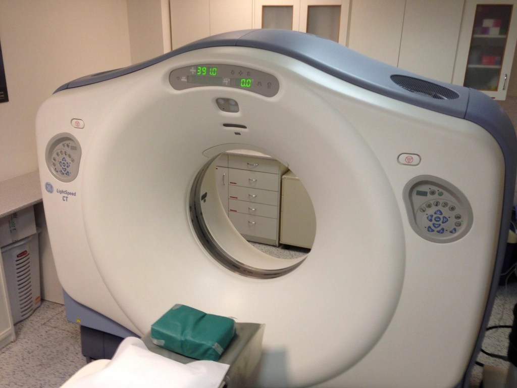
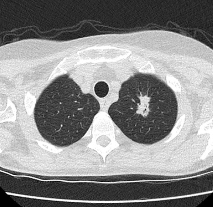
Computed Tomography (CT) scans are fast scans of the body with X rays. It will feel like you are moving through the donut over the course of seconds. A 3D image will be able to show doctors high resolution images of your bones and organs. Sometimes, iodinated contrast is injected into your veins prior to the scan to better show blood vessels and certain organs. Tumors tend to have leaky disorganized blood vessels because they grew in such a hurry, so contrast will leak through and show some tumors better.

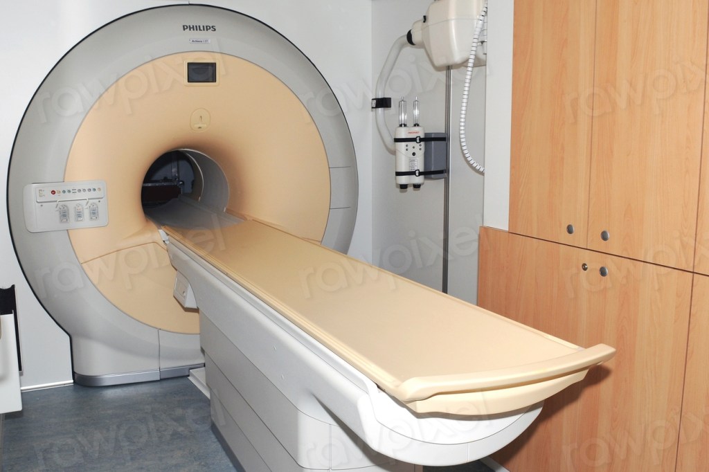
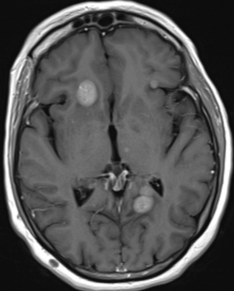
Magnetic Resonance Imaging (MRI) scans can take 20 minutes to an hour, depending on the area being scanned and how many different images the doctors want to see. As the name indicates, there is a large magnet that forces all the water molecules in your body to be aligned to the same field. Then loud bursts of radiofrequency energy are used to disrupt the alignment in the area being scanned (you will be given ear plugs). As the water molecules re-align back to the magnetic field, they release radiowaves that are detected to make an image. Muscle, bone, body fluids, and fat all give different signals and we can get better contrast with MRI than CT scans. MRIs are most often used to image the brain, spinal cord, or abdomen where CT can’t distinguish all the different organs as well.
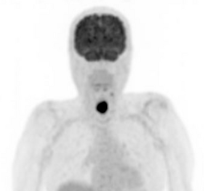
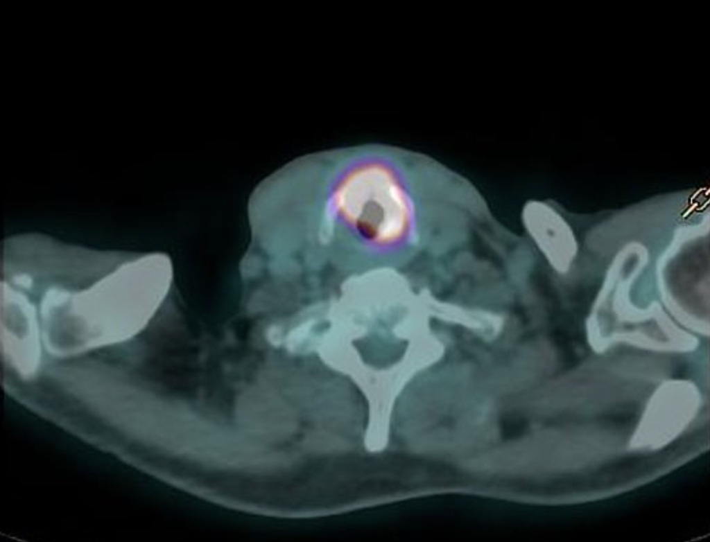
Positron emission tomography (PET) typically involves injecting a radioactive sugar solution (FDG) into your veins, which is absorbed by cancer cells because they are actively using sugar metabolism to grow. Other normal organs such as the brain will light up because those cells also preferentially use sugar for its energy source. After some time to allow the sugar to be taken up, you will be scanned in a combined PET and CT scanner. The low level radiation from FDG will be detected by the scanner and overlaid on the CT scan. This scan is useful because it is more sensitive than a CT scan for detecting cancer metastasis/spread.
Biopsy and Pathology
The ultimate step to diagnosis requires taking a sample from the tumor, usually through a needle, and then looking at its characteristics under a microscope. Cancer cells typically look disorganized, have large amounts of DNA replicated in them, are in the middle of dividing, and ultimately can be seen invading through normal barriers or may even be seen invading lymph/blood vessels or nerves. The pathologist will also stain the cells for different markers to determine the origin of the cancer cell. Some markers are important for making treatment decisions, such as if breast cancer expresses estrogen receptor on the surface. Others may tell us how aggressive the cancer is acting (eg. ki-67). There is active research trying to identify genetic and immune alterations to your cancer that we have drugs to target.

Once your doctors know what type of cancer you have and how far it has spread, then they can come up with a treatment plan with you.
